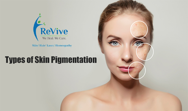
Pigmentation is nothing but the coloration of the skin, and melanin is the pigment responsible for skin color. The lesser the melanin the body produces, the lighter the skin and vice versa. There are many kinds of skin pigmentation, like
- Freckles: These are the most common type of skin pigmentation. They are nothing but tiny closely-held dots caused by excessive exposure to the sun. They develop into darker spots after repeated exposure. If you have a fair complexion, your freckles will be more visible.
- Melasma: The pigmentation condition is prevalent in women. These are generally large tan or brown patches that appear on the face. They are triggered by changes in hormones and can get darker with sun exposure, medications, stress, and even pregnancy. That is why they are often called, Pregnancy marks.
- Vitiligo: It is another pigmentation condition that causes smooth white patches on the skin. Vitiligo happens due to a lack of pigment-producing cells called melanocytes in the skin. This is why skin appears lighter than normal. These patches can be very sensitive to the skin, and this disorder has no set cure by far.
- Lentigines: These are also called liver spots. These are the pigmentation spots that are dark in color. They are irregular in shape and occur due to the presence of high amounts of melanocytes in the superficial layers of the skin. This condition is commonly seen in middle age and elderly part of the population. The face and the back of your hands are the most affected in this condition.
- Post-Inflammatory Hyperpigmentation: This type of skin pigmentation is caused due to acne or sunburn. One thing about this kind of pigmentation is that it can be reversed with topical products.
- Peri-orbital Melanosis: This skin discoloration is commonly known as dark circles. A person with this condition usually inherits it, but it can result from stress or strain on the eyes.
- Photo-melanosis: A condition that results from sun exposure and is characterized by patches of pigmentation on the face, neck, back, and arms.
- Sunburn: A type of reddish discoloration of the skin caused by too much exposure to the sun. This type of discoloration is most common in people with fairer complexions.
- Albinism: Albinism is an inherited disorder caused by the absence of the melanin pigment. This condition results in no pigmentation in the skin, hair, or eyes. Albinism is caused by a gene abnormality that causes melanin production to be limited. The condition has not yet been cured. Those with this disorder should always use sunscreen since sun damage and skin cancer are more likely to occur.
Many people are affected by birthmarks and other pigmentation disorders. This abnormal skin coloration is likely to appear at birth or within a few weeks after birth. Most birthmarks are noncancerous, but certain birthmarks, described below, can pose health risks. There are different birthmarks, such as pigmented, macular stains, hemangiomas, and port-wine stains. The various birthmarks are discussed below:
- Nevus of Ota: This birthmark is characterized by bluish or grayish discoloration of the face and the white part of the eye (sclera). The increased amounts of the pigment called melanin and the cells that produce this pigment (melanocytes) in and around the eyes cause discoloration. This type of birthmark increases patients’ risk of melanoma of the eye or central nervous system. They may also develop Glaucoma. As a result, neurologists and ophthalmologists should examine them regularly.
- Mongolian spots. The birthmarks usually appear bruised or bluish and typically appear on the back or buttocks of babies. The discoloration usually disappears by the age of four and needs no treatment.
- Cafe-au-lait spots: These are light brown-to-dark brown flat spots with smooth or irregular borders. Approximately 10% of the general population has 1 or 2 spots and does not have any other disorder associated with them. However, several of these spots larger than 0.5 cm can be a sign of neurofibromatosis.
- Nevi (moles): These spots are usually skin-colored to light or dark brown and flat or raised. Moles are most commonly benign (noncancerous) and do not cause any problems. There is a possibility that some moles may turn into melanoma. Therefore, monitoring a mole for bleeding, pain, itching, color, symmetry, and even borders is important.
Now, there are a few vascular birthmarks. Some of them are discussed below.
- Macula: The macula is a mild red mark that appears anywhere on the body but is not elevated. Macular birthmarks are among the most common types. It can occur as angel kisses or stork bites, which appear on the back of the neck and last into adulthood. Angel kisses can appear on the forehead and eyelids and usually disappear after age two. Stork bites will appear on the back of the neck and last into adulthood. Usually mild and harmless, these marks don’t need to be treated.
- Hemangioma: Hemangiomas are masses of blood vessels that are tightly clustered together. There are some more severe hemangiomas. Premature babies and females are more likely to get them. It usually manifests as a small spot on the face, the trunk, or the extremities. Hemangiomas, however, can be large and grow rapidly in some children during the first year of life.
- Port-wine stains: Port-wine stains appear as flat spots of pink, red, or purple on the face, trunk, arms, or legs and are permanent. These stains occur due to abnormal blood vessel development (capillaries). The port-wine stain can become raised and thickened over time. The presence of port-wine stains on the eyelids increases the risk of Glaucoma. Port-wine stains may appear in certain medical disorders, such as Sturge-Weber Syndrome. Port-wine stains on the face are associated with vision problems, convulsions, intellectual disabilities, and paralysis. Another is Klippel-Trenaunay Syndrome, in which port wine stains, varicose veins, and excessive bone growth may appear in the limb. The two syndromes are sporadic.